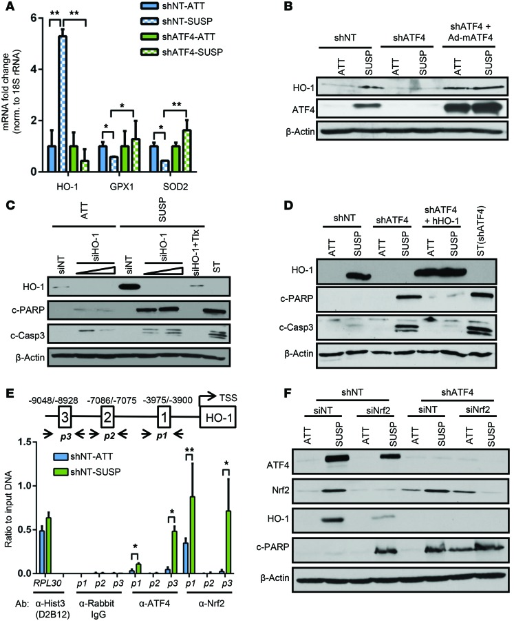
Figure 5. ATF4 induces expression of HO-1 in response to detachment-activated oxidative stress.
(A) RT-PCR analysis of HO1, GPX1, and SOD2 in shNT and shATF4.HT1080 cells grown in suspension for 48 hours. Data are represented as mean fold change compared with attached cultures for 3 independent experiments (n = 3, mean ± SD). *P < 0.05; **P < 0.01, Student’s t test. (B) Expression of HO-1 in shNT and shATF4.HT1080 cells and adeno-mATF4–infected shATF4.HT1080 cells as measured by immunoblot analysis. (C) HT1080 cells were transfected with increasing amounts of siRNA against HO-1 (siHO-1) or siNT. Immunoblots for indicated proteins were measured. (D) shATF4.HT1080 cells with lentiviral-mediated overexpression of hHO-1 (shATF4 + hHO-1) were grown. Immunoblots for apoptosis markers were performed. (E) Schematic representation of AREs in the HO1 promoter and primer sets (p1, p2, p3) indicating amplified regions encompassing the 3 ARE sites along with transcription start site (TSS). ChIP analysis was performed by either α-ATF4 or α-NRF2 antibody and by measuring enrichment at 3 AREs in human HO1 promoter by RT-PCR. Antibodies against histone 3 (α-histone 3 D2B12) and rabbit IgG (α-rabbit IgG) were used as positive and negative controls, respectively, to amplify human RPL30 exon 3 of the human genome. Data are represented as ratio of input DNA (1:25) and presented as mean of 2 independent experiments (n = 2, mean ± SD). *P < 0.05; **P < 0.01, Student’s t test. (F) Immunoblot analysis for indicated proteins in shNT and shATF4.HT1080 cells transfected with siNT or siNRF2 after 48 hours of suspension. All immunoblots are representative of 2 independent experiments.

