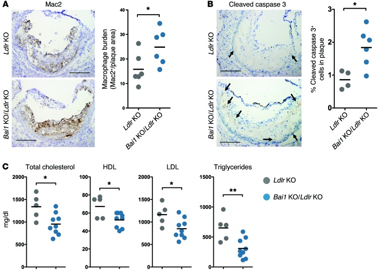
Figure 5. BAI1 signaling affects serum lipid levels in dyslipidemic Ldlr-deficient mice.
(A) Immunohistochemistry for the macrophage marker Mac2 within the aortic roots of Bai1 KO/Ldlr KO mice and control Ldlr–/– littermates after 15 weeks on a Western diet. Quantification of Mac2+ area normalized to oil red O+ area (Supplemental Figure 6D) (both measured using ImageJ software) as an indication of macrophage burden is shown on right. *P < 0.05 (t test). (B) Immunohistochemistry for cleaved caspase 3 (indicative of cells undergoing apoptosis) within the aortic roots of Bai1 KO/Ldlr KO mice and control Ldlr–/– littermates after 15 weeks on Western diet. Scale bars: 200 μm. Graph represents the number of cleaved caspase 3–positive cells (arrows) counted in the atherosclerotic plaques of each mouse in a representative cohort (of 2 independent experiments producing similar results). *P < 0.05 (t test). (C) Analysis of serum levels of total cholesterol, HDL, LDL, and triglycerides in Ldlr–/– mice compared with Bai1 KO/Ldlr KO mice after 15 weeks on a Western diet. *P < 0.05; **P < 0.01 (t test).

