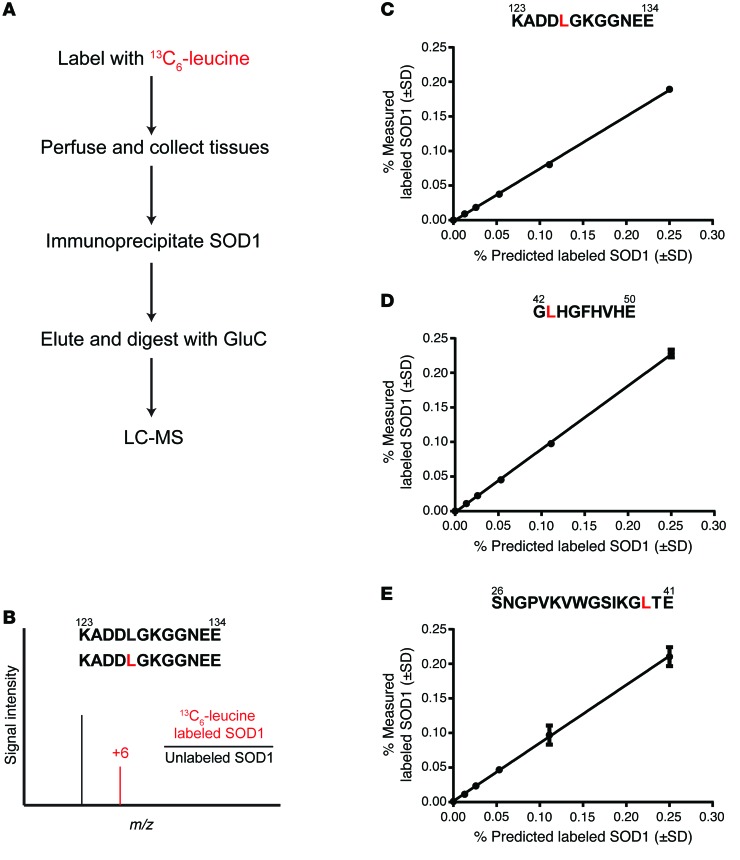
Figure 1. Schematic of SOD1 isolation and mass spectrometry detection method.
(A) Flow chart detailing the isolation, processing, and detection of leucine-containing SOD1 peptides. GluC, endoproteinase Glu-C; LC-MS, LC/tandem MS. (B) Schematic representing LC/tandem MS detection of 13C6-leucine–containing peptides. The +6-Da shift in the leucine-containing peptide KADDLGKGGNEE reflects the incorporation of 13C6-leucine. (C–E) LC/tandem MS standard curves for the 3 leucine-containing peptides used in quantifying labeled SOD1.

