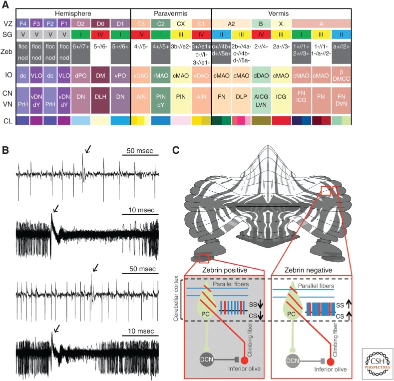
Figure 3.
Olivocerebellar modules and Purkinje cell activity in relation to zebrin (II) distribution. (A) The VZ and SG rows refer to the zones and groups of Purkinje cells described by Voogd (1964; VZ) and Sugihara (2011; SG), respectively. The Zeb row indicates which zones and groups are zebrin positive (grey) and zebrin negative (white). The inferior olive (IO) row indicates which subnucleus of the IO is providing climbing fibers to a particular zone/group of Purkinje cells in the cerebellar cortex and collaterals to a particular part of the cerebellar nucleus, depicted in the same column. The CN and VN row indicates the parts of the cerebellar nuclei (CN) and vestibular nuclei (VN) that are innervated by the strip of Purkinje cells, depicted in the same column. It should be noted that only those vestibular nuclei are indicated that both receive a Purkinje cell input and provide a feedback projection to the inferior olive; because, for example, medial and superior vestibular nuclei do not project to the IO, they are not incorporated in this scheme. In addition, it should be noted that this overview is also incomplete in that some nuclei, such as the dorsomedial cell column (DMCC), may receive inhibitory feedback from multiple hindbrain regions. CL indicates the color legends used for Figure 2. AICG, anterior interstitial cell group; AIN, anterior interposed nucleus; β, subnucleus β; c/rMAO, caudal/rostral medial accessory olive; dc, dorsal cap; DLH, dorsolateral hump; DLP, dorsolateral protuberance; DM, dorsomedial group; DMC, dorsomedial crest; d/vDAO, dorsal/ventral dorsal accessory olive; DVN, descending vestibular nucleus; d/vPO, dorsal/ventral principal olive; dY, dorsal group Y; floc, flocculus; FN, fastigial nucleus; ICG, interstitial cell group; LVN, lateral vestibular nucleus; nod, nodulus; PIN, posterior interposed nucleus; PrH, prepositus hypoglossal nucleus; vDN, ventral dentate nucleus; and VLO, ventrolateral outgrowth. (B) Examples of raw traces of Purkinje cell activity from zebrin-positive (top panels) and zebrin-negative (bottom panels) zones. Arrows indicate complex spike. (From Zhou et al. 2014; reprinted, with permission, from the authors.) (C) Intramodular connections via deep cerebellar nuclei (DCN) explaining why the complex spike (CS) activity within a module follows the intrinsic differences in simple spike (SS) activity of Purkinje cells (PC). PC and DCN are inhibitory, whereas climbing fibers are excitatory. (From Albergaria and Carey 2014; reprinted under the terms of the Creative Commons Attribution License, which permits unrestricted use and redistribution provided that the original author and source are credited.)

