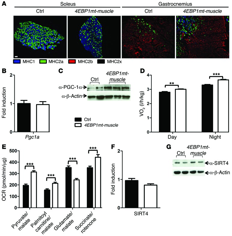
Figure 2. Tg-4EBP1mt-muscle mice exhibit muscle type transformation and increased oxygen consumption.
(A) Immunofluorescence staining of type I skeletal muscle fibers (MHCI, blue) and type II skeletal muscle fibers (MHC2a, green; MHC2b, red; and MHC2x, unstained). Scale bar: 100 μm. (B) RT-PCR quantification of Pgc1a expression in quadriceps normalized to Ppia expression (n = 4). (C) Western blot analysis of PGC-1α expression in quadriceps. (D) Oxygen consumption measured in a 3-day/night period in 6-month-old male mice fed a normal chow diet (n = 8–12 per genotype). (E) State III oxygen consumption rates (OCRs) from isolated skeletal muscle mitochondria in the presence of indicated substrates were measured in an XF24 Extracellular Flux Analyzer (n = 5 per genotype). (F) RT-PCR and (G) Western blot analysis of SIRT4 expression in quadriceps. P values were assessed by a 2-way ANOVA. Bonferroni post-tests were used to compare replicate means by row. **P < 0.01; ***P < 0.001. RT-PCR samples were from n = 4 per genotype.

