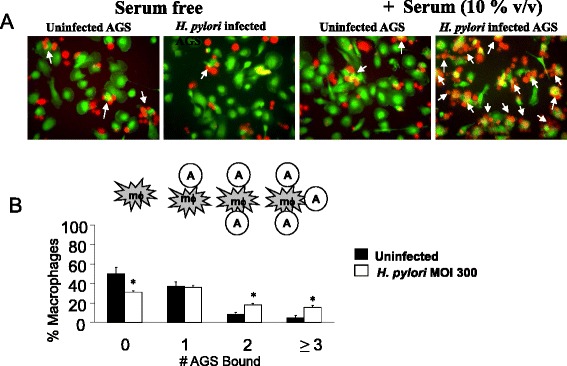
Fig. 1.

Infection of AGS cells with H. pylori increases interactions with macrophages and these interactions are facilitated by C1q. a The association of macrophages (green) and AGS cells (red) is shown in the absence and presence of serum, with either uninfected AGS cells or AGS cells that had been infected with H. pylori (MOI 300) for 24 h. Macrophages with aggregates of attached AGS cells are marked with a white arrow. b THP-1 macrophages were incubated for 1 h with either uninfected AGS (black) or H. pylori-infected (MOI 300) AGS (white) cells after which time the slide was washed three times and the number of bound AGS cells was counted in four random fields. Data shown is the average of n = 5–7 independent experiments. * overall P value of < 0.05
