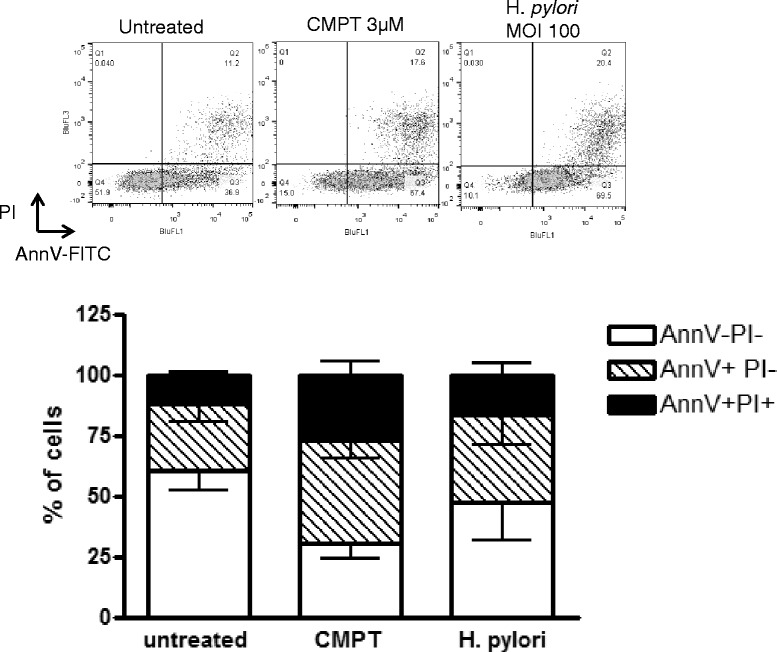
Fig. 3.

Induction of apoptosis in AGS cells. Induction of apoptosis in AGS cells by camptothecin (3 μM; 24 h) and H. pylori (MOI 100; 24 h). The top panel shows representative dot plots of AGS cells stained with Annexin V-FITC and PI in control untreated cells (left), following camptothecin treatment (middle) and H. pylori treatments (right). The bottom graph shows cumulative data from four independent experiments. AnnV-PI- are considered healthy, AnnV + PI- are considered to be apoptotic and AnnV + PI+ are cells which have progressed to secondary necrosis
