Figure 7.
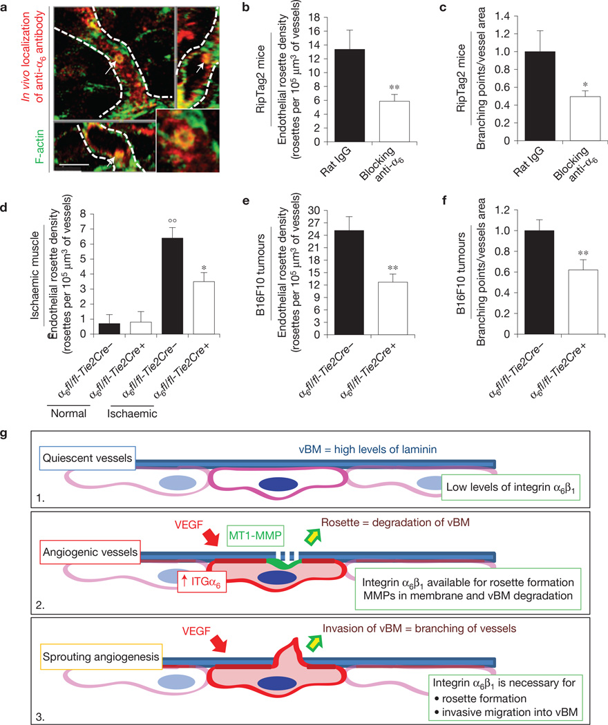
In vivo blocking of integrin α6 impairs endothelial podosome rosette formation and reduces vessel branching in tumours. (a) Rapid accumulation of anti-α6 integrin antibody into endothelial podosome rosettes of RipTag2 tumour vessels. xyz-sections of confocal micrographs of the distribution of immunoreactivity in RipTag2 tumours 10 min after intravenous injection of 25 µg of anti-α6 integrin antibody. Vessels are delimited by white dotted lines; white arrows indicate the podosome rosette. Inset, high magnification of the podosome rosette. Scale bar, 5 µm. (b) Measurements of rosette density in vessels of RipTag2 mouse tumours, treated with rat IgG or anti-α6 blocking antibody. Mean ± s.e.m. of n = 30 fields, 5 fields per pancreatic islet from 6 mice per treatment group. Statistical significance was calculated using an unpaired non-parametric Mann-Whitney test (**P<0.01 versus rat IgG.). (c) Branching density in blocking antiα6-treated RipTag2 tumours. Mean ± s.e.m. of n = 30 fields, 5 fields per mouse from 6 mice per treatment group. Statistical significance was calculated using an unpaired non-parametric Mann-Whitney test (*P<0.05 versus rat IgG). (d) Measurements of rosette density in vessels of gastrocnemius muscles from unilateral hindlimb ischaemia experiments in WT (α6fl/fl-Tie2Cre−) or endothelial α6 null (α6fl/fl-Tie2Cre+) mice. Mean ± s.e.m. of n = 9 fields, 3 fields per muscle from 3 mice. Statistical significance was calculated using an unpaired non-parametric Mann-Whitney test (°°P < 0.01 versus normal α6fl/fl-Tie2Cre−; *P<0.05 versus α6fl/fl-Tie2Cre+). (e) Measurements of rosette density in vessels of subcutaneous B16-F10 tumours in WT (α6fl/fl-Tie2Cre−) or endothelial α6 null (α6fl/fl-Tie2Cre+) mice. Mean ± s.e.m. of n = 21 fields, 3 fields per tumour from 7 mice per treatment group. Statistical significance was calculated using an unpaired non-parametric Mann-Whitney test (**P< 0.01 versus α6fl/fl-Tie2Cre−). (f) Branching density in B16F10 melanoma subcutaneously injected in Tie2-dependent α6 KO mice. Mean ± s.e.m. of n = 42 fields, 5 fields per tumour from 7 mice per treatment group. Statistical significance was calculated using an unpaired non-parametric Mann-Whitney test (**P<0.01 versus α6fl/fl-Tie2Cre− mice). (g) Cartoon showing α6 integrin/laminin molecular mechanisms involved in sprouting angiogenesis. (1) Quiescent EC have low levels of α6β1 integrin, which binds vBM laminin, is recruited in FAs, and results in blood vessel stabilization. (2) When the tumour produces VEGF, the VEGF induces upregulation of the α6 integrin subunit in ECs. The increased availability of α6β1 integrin then allows the formation and stabilization of endothelial podosome rosettes and the ensuing MMP-driven degradation of ECM that, in turn, (3) allows vBM invasion by ECs and sprouting angiogenesis.

