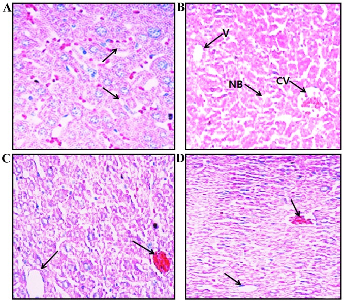
Figure 2.
Histopathological examination of mouse liver tissue stained with H&E at ×20 magnification. (A) Control mice (group I), central vein and normal hepatocytes are evident. (B) AA-treated mice (group II), cytoplasmic vacuolation, cell necrosis, central vein blood deposition and chronic periportal inflammatory cell infiltration are present. (C) AA + MH5-treated mice (group III) and (D) AA + MH15-treated mice (group IV), AA-induced steatosis and other pathological changes were reduced. Necrotic bodies (NB), cental veins (CV) and vacuoles (V) are indicated by black arrows. Images were captured using a light microscope at ×20 magnification. AA, acrylamide; AA + MH5, acrylamide 50 mg/kg plus morin hydrate 5 mg/kg; AA + MH15, acrylamide 50 mg/kg plus morin hydrate 15 mg/kg.

