
Figure 2.
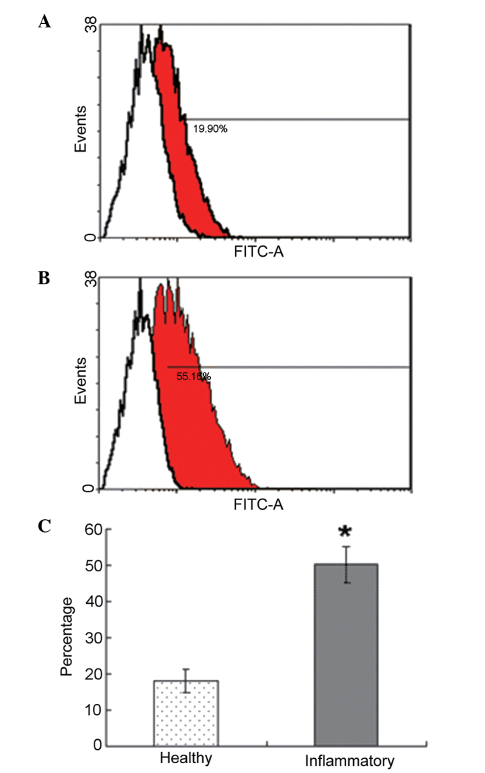
Periodontitis-associated changes in the number of PDLSCs. (A) Flow cytometry data of PDLCs from a healthy donor. (B) Flow cytometry data of PDLCs from a periodontitis-affected donor. (A and B) Red areas indicate STRO-1-positive cells, white areas indicate STRO-1-negative cells. (C) Numbers of PDL-derived STRO-1+ cells were significantly increased in the inflammatory group, compared with the healthy group (*P<0.05). PDLC, periodontal ligament cell; FITC, fluorescein isothiocyanate.