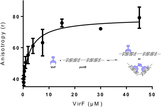
Fig 5. Plot of MalE-VirF Binding to the pvirB DNA Probe.
For the assay, 5’Fluoroscein-pvirB DNA probe concentration was held constant at 50 nM, while MalE- VirF concentration was varied from 45 μM to 0.12 μM. The observed binding max for MalE-VirF binding was approximately r = 75, while the observed baseline (no MalE-VirF, only free pvirB DNA probe) was r = 42. The assay was conducted in duplicate. The inset depicts the reaction being monitored, VirF (not known if monomer or dimer) binding to the virB promoter.

