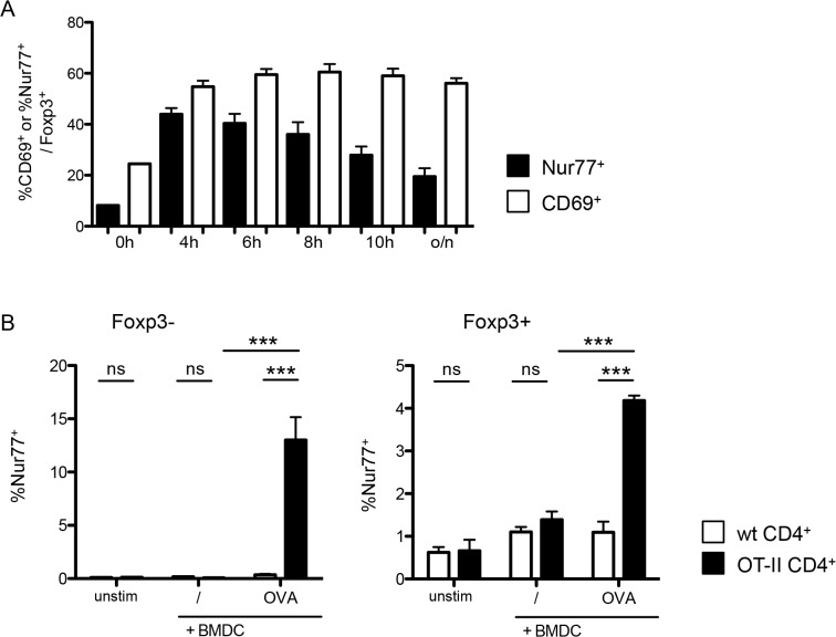
Fig 3. Foxp3+ cells upregulate Nur77 after TCR activation in an antigen-specific manner.
(A) Time course of Nur77 (black bars) and CD69 expression (white bars) on Foxp3+ T-cells among sorted CD25+ CD4+ splenocytes stimulated with plate-bound αCD3 and 1U/ml IL-2. Data are representative of 3 independent experiments with n ≥ 3. (B) Nur77 expression on Foxp3- (left) and Foxp3+ (right) among enriched CD4+ T-cells from either wild-type (white bars) or OT-II (black bars) mice, co-cultured overnight with unstimulated, OVA pulsed or αCD3 coated BMDCs. Data are representative of two independent experiments, with each n = 3 per group. unstim, control without BMDC; /, unpulsed BMDC; OVA, OVA-pulsed BMDC, anti-CD3, anti-CD3-coated BMDC. Data show mean + SD, *p ≤ 0.05, *** p ≤ 0.001; ns, non significant. Data are representative of four independent experiments each with n = 3 per group.

