
Figure 6.
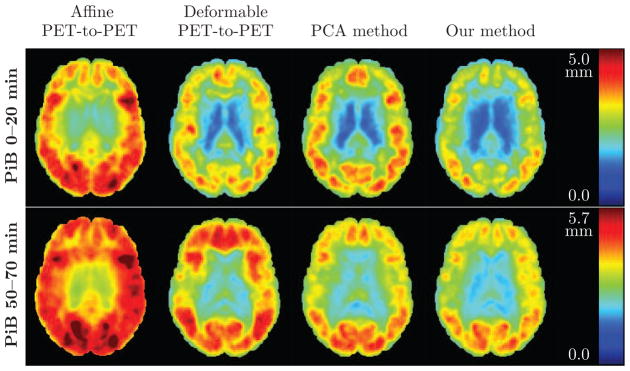
Root mean square (RMS) error (in mm) of the PET deformation fields obtained from different approaches, calculated across 79 subjects. An axial brain slice is shown. Top row: Results for 0–20 minute PiB-PET. Bottom row: Results for 50–70 minute PiB-PET. Left to right: RMS error of the affine PET-to-PET registration T′, RMS error of the PET-to-PET deformation ψ, RMS error of the deformation given by the PCA method, and RMS error of ξ̂ predicted using our model. For references to color, the reader is referred to the web version of this article.