
Figure A.1.
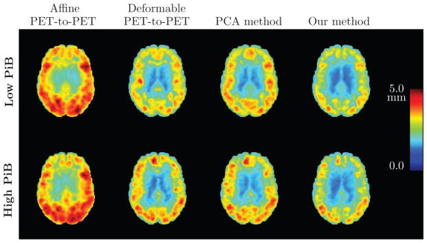
Root mean square (RMS) error (in mm) of the PET deformation fields obtained from different approaches based on 0–20 minute PiB-PET registration, calculated across 79 subjects. An axial brain slice is shown. Top row: Results for low PiB individuals. Bottom row: Results for high PiB individuals. Left to right: RMS error of the affine PET-to-PET registration T′, RMS error of the PET-to-PET deformation ψ, RMS error of the deformation given by the PCA method, and RMS error of ξ̂ predicted using our model.