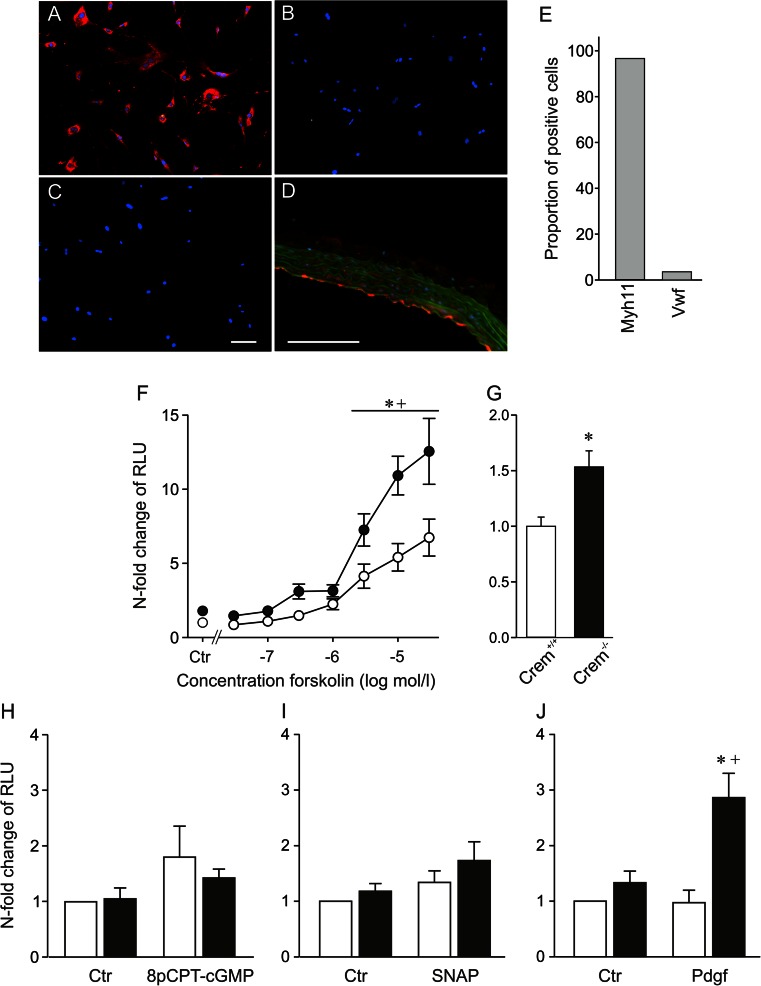
Fig. 1.
Characterization of VSMC culture by immunofluorescence staining (a–e). Photographs of isolated VSMCs incubated with an antibody against the a smooth muscle myosin heavy chain (Myh11), b Von Willebrand factor (Vwf), c the secondary antibody control without primary antibody, and d positive control of the Vwf antibody in aortic sections; note the positive red staining of the endothelium. e Quantification of the proportion of stained cells based on 350 analyzed cells revealed over 95 % positive staining of the cells by the VSMC marker Myh11; scale bar 100 μm. f–j CRE-mediated transcriptional activation of isolated VSMCs from Crem+/+ (white symbols) and Crem−/− (black symbols) mice. VSMCs were transiently transfected with a CRE-controlled luciferase reporter gene construct and treated with activators of different signaling cascades or solvent controls (Ctr). f Forskolin (in DMSO, n = 8/6 meaning eight independent transfections from six isolations, for 12 h; Ctr: n = 15–18/6), h 10−4 M 8-pCPT-cGMP (in H2O, n = 6/6, for 9 h), i 10−4 M SNAP (in H2O, n = 7/7, for 9 h), and j 7.5 ng/ml Pdgf (in H2O, n = 5/5, for 9 h). Data were presented relative to the mean of Crem+/+ Ctr, which was set to 1. Note the elevated CRE-dependent transcriptional activity under stimulation with 3 × 10⁻⁸–3 × 10⁻⁵ M Forskolin (f) and Pdgf (j) in VSMCs of Crem−/− mice. Independent analysis of untreated transfected VSMCs (g) revealed a 1.5-fold increase in VSMCs from Crem−/− vs. Crem+/+ mice (n = 51–54/3). Asterisk (*) denotes p < 0.05 vs. Crem+/+, a cross (+) denotes p < 0.05 vs. control

