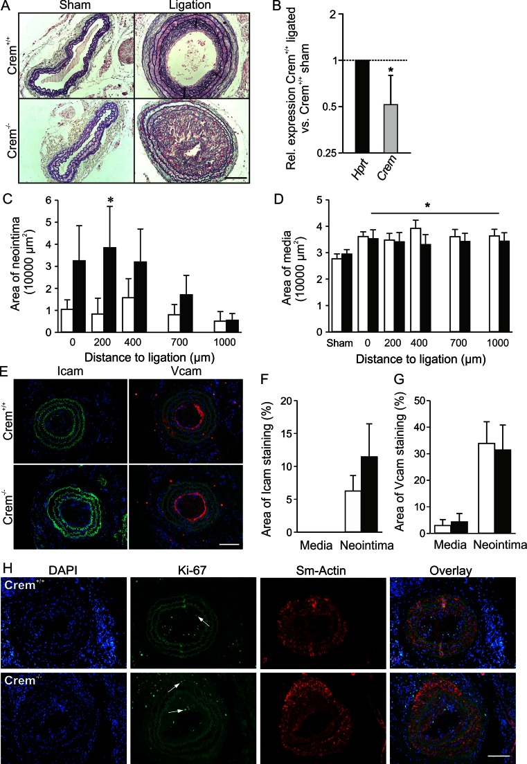
Fig. 4.
Analysis of neointima formation of the right carotid artery 3 weeks after ligation. a Resorcin-fuchsine and nuclear fast red staining of the cross sections of the ligated right and sham-operated left carotid artery from Crem+/+ and Crem−/− mice. b Downregulation of relative Crem mRNA levels in Crem+/+ sham (n = 6) and ligated carotids (n = 4). Quantification of neointima (c) and media area (d) in Crem+/+ (white, n = 10) and Crem−/− mice (black, n = 9). Note the severe increase in neointima formation in the Crem−/− mice. e Icam1- and Vcam1-specific immunofluorescent staining of carotid cross sections of ligated carotids from Crem+/+ and Crem−/− mice. Quantification of Icam1- (f) and Vcam1 (g)-stained area showed no differences between the genotypes. *p < 0.05 vs. Crem+/+ (post hoc tests, two-way ANOVA). h Detection of proliferating VSMCs in the cross sections of ligated carotids. Photomicrographs show the following: the cell nuclei stained with DAPI, proliferating cells (VSMCs) detected by a Ki-67 antibody (white arrows), visualization of VSMCs with a smooth muscle actin-specific antibody, and the overlay. Crem−/− VSMCs showed an increase in the percentage of proliferating VSMCs in the media but not in the neointima (for details, see text). Scale bars = 100 μm

