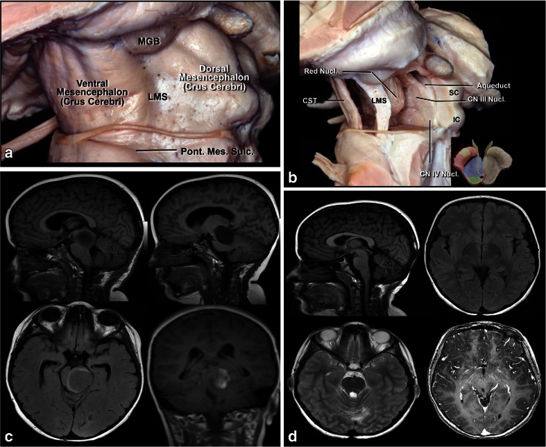
Fig. 8.
a Lateral view of the midbrain. The lateral mesencephalic sulcus (LMS) runs on the surface of the midbrain extending from the medial geniculate body (MGB) above to the pontomesencephalic sulcus (Pont. Mes. Sulc.) below. The LMS extends along the lateral edge of the medial lemniscus (ML). b The ML divides the midbrain into ventral (anterior) and dorsal (posterior) parts. Neurocritical structures at entry through the LMS are the corticospinal tract in the anterior midbrain, the red, oculomotor and trochlear nuclei in the central (tegmentum) midbrain, nuclei of the superior and inferior colliculus in the posterior (dorsal) midbrain. The oculomotor nucleus is positioned at the level of the inferior half of the superior colliculus and superior half of the inferior colliculus in the midline, and the trochlear nucleus is positioned at the level of the inferior half of the inferior colliculus in the midline. c Lateral lesion in the central midbrain approached via infratentorial supracerebellar route along the lateral sulcus of the mesencephalon. d Five-year follow-up after complete resection showing no evidence of the lesion, a pilocytic astrocytoma

