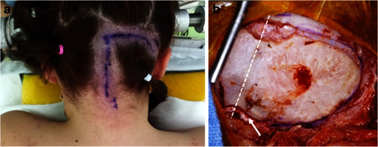
Fig. 1.
Posterior fossa craniotomy for CPA tumors. a A hockey stick incision with a midline incision from C2 spinous process level with an upper lateral incision extending toward the base of mastoid process. b A surgical photograph showing a posterior fossa craniotomy of bilateral occipital bone with the ipsilateral side wider, extending to just medial to the sigmoid sinus. A dashed line indicates the sagittal midline. Note the craniotomy crosses the foramen magnum (arrow)

