Fig. 9.
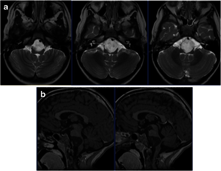
Case 3. Four-year-old girl with previous history of chemical meningitis. MR images (a T2-weighted axial view, b T1-weight sagittal image after contrast infusion) showing a large mass ventral to the ponto-medullary junction. Note a severe brain stem compression by the non-enhancing tumor between the brain stem and the basilar artery

