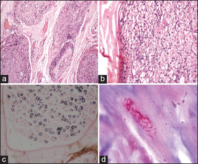
Figure 2.

Biopsy features of borderline lepromatous leprosy PNL. (a) The cross-section of the nerve showing minimal endoneurial inflammation (H and E ×40). (b) The endoneurium showing foam cells (H and E ×100). (c) Kpal stain showing reduction in the number of myelinated fibers with few thinly myelinated fibers (arrowheads) (Kpal ×100). (d) Ziehl Neelsen (5%) stain showing lepra bacilli (arrow)
