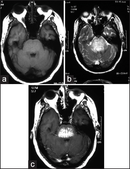
Figure 1.

(a): Axial T1-weighted MRI showing a bulky pons with an isointense lesion (b) The lesion was hyperintense on T2-weighted MRI (c) Contrast MRI with gadolinium showed multiple punctuate enhancing lesions giving the classic “peppering of pons” appearance
