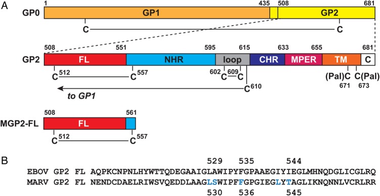
Figure 1.

Schematic of the Marburg virus (MARV) GP2 fusion loop (FL). A, The MARV glycoprotein is produced as a single precursor (GP0) that is cleaved by furin during maturation into the 2 subunits GP1 and GP2. The primary sequence of GP2 contains the FL, N-heptad repeat region (NHR), loop, C-heptad repeat region (CHR), membrane proximal external region (MPER), transmembrane domain (TM; which includes 2 palmitoylated cysteines, as indicated), and C-terminal tail (C). The GP1 subunit is disulfide bonded to the GP2 subunit via an intramolecular disulfide bond from C610. The Marburg virus GP2 fusion loop (MGP2-FL) protein studied here consists of residues 508–561, with the native internal disulfide bond between C512 and C557. B, Amino acid sequence alignment between the EBOV GP2 FL and the MARV GP2 FL (note that residue numbering differs by 1 position). The positions of residues that form the core of the EBOV GP2 FL hydrophobic fist [9] are indicated. The aligning positions in MARV glycoprotein FL, as well as 2 others (all indicated in blue), were subjected to alanine scanning mutagenesis. Abbreviation: EBOV, Ebola virus.
