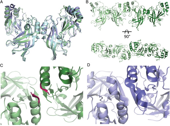
Figure 2.
A, Alignment of VP40 dimers from Sudan virus (SUDV) and Ebola virus (EBOV). SUDV (Protein Data Bank [PDB] 3TCQ) is shown in green, and two EBOV structures in blue (PDB 1ES6) and cyan (PDB 4LDB). VP40 dimers are thought to further oligomerize via the conserved C-terminal domain (CTD)-to-CTD interface. B, CTD-to-CTD interface between two SUDV VP40 dimers is shown in two orientations. C, Residues 232–234 were not visible in a lower-resolution structure of SUDV VP40 (PDB 4LD8), but they are clearly visible in this crystal structure. They contribute to the CTD-to-CTD interface and are shown in magenta. D, Same interface in EBOV is shown for comparison.

