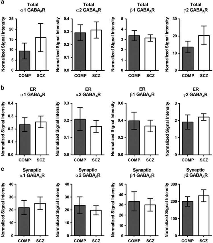
Figure 4.
α1, α2, β1 and γ2 GABAAR subunit expression are not different between diagnostic groups in the total homogenate, ER or SYN fractions. Western blot analysis of α1, α2ALL, β1 and γ2 GABAAR subunit expression in schizophrenia and comparison subjects. There are no differences between diagnostic groups in the protein expression of α1, α2ALL, β1 or γ2 GABAAR subunits in (a) the total homogenates, (b) ER fractions or (c) the SYN fractions. Data are expressed as the mean signal intensity (±s.e.m.) of protein targets in the ER fraction normalized to VCP as a loading control, and JM4 as an ER marker or gephyrin as an inhibitory synapse marker, relative to the VCP-normalized signal intensity of the same target in the total homogenate. COMP, comparison subject; ER, endoplasmic reticulum; GABAAR, γ-aminobutyric acid type A receptor; SCZ, schizophrenia; SYN, synapse; VCP, valosin-containing protein.

