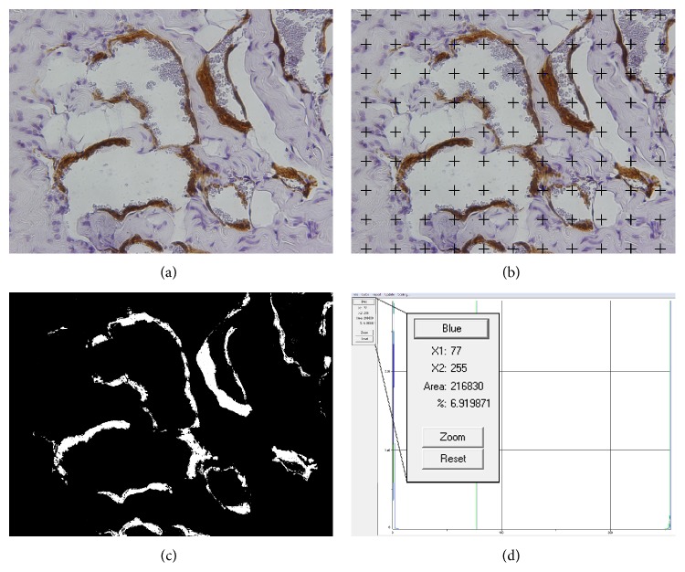
Figure 1.
(a) Example of a histological field of a rat's corpus cavernosum immunostained with antismooth muscle α-actin and captured under a ×400 magnification field. (b) The same field after superimposition of the 99-point grid. The points touching the smooth muscle were counted. (c) The same field after all smooth muscle areas was transformed into white colored pixels while the remaining pixels of the images appear in black. (d) Histogram data of image (c) showing that 6.9% of the image is composed by white pixels, that is, smooth muscle.

