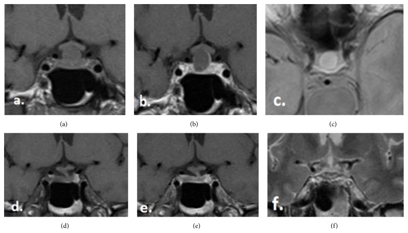
Figure 1.
(a–c) The MRI images by the initial diagnosis, (a) T1-coronal without contrast, (b) T1-coronal with contrast, and (c) T2-axial without contrast. (d–f) The MRI images by the 1-year follow-up, (d) T1-coronal without contrast, (e) T1-coronal with contrast, and (f) T2-coronal without contrast.

