
Figure 3.
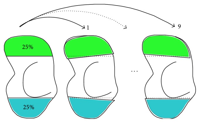
Scheme of the cadaveric experiment assessing the accuracy and reproducibility of the matching procedure. After scanning one cadaveric arm tenfold, the scaphoid from one CT scan was segmented (left model). The proximal (blue) poles were matched to the remaining 9 scans enabling displacement analysis of the distal (green) pole.