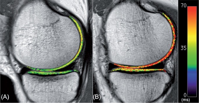
Figure 2.

Color-coded sagittal T2 maps in the medial compartment of the contralateral knee (A) and the ACL-reconstructed knee (B) of one patient. Note the elevated T2 values of articular cartilage in the femoral compartment of the ACL-reconstructed knee relative to the contralateral knee.
