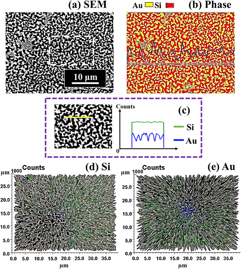
Figure 5. Elemental analysis of self-assembled Au nanostructures on 4H-SiC (0001) by the energy-dispersive X-ray spectroscopy (EDS).
(a) SEM image of the sample with the 8 nm DA annealed at 600 °C. (b) Combined EDS phase map of Au (yellow) and Si (red). (c) Line-profiles of element counts of Si (green) and Au (blue) denoted with the yellow line in the SEM image. (d,e) 3-D top-view phase maps of Si and Au.

