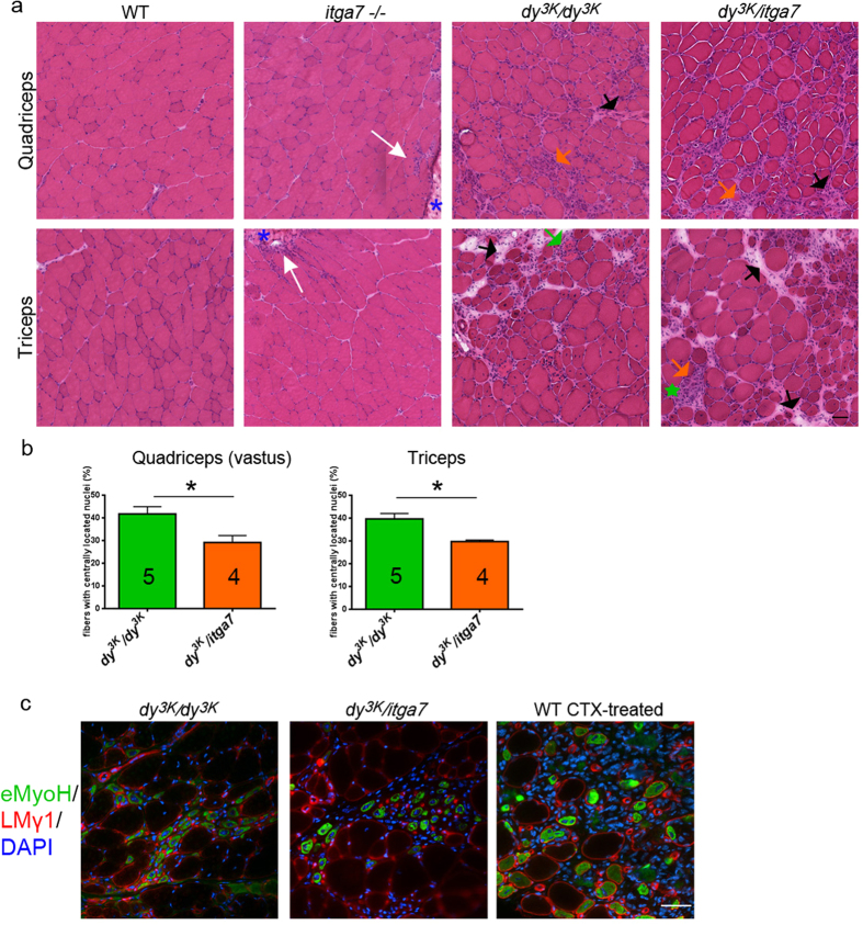
Figure 3. Muscular dystrophy hallmarks are not exacerbated in limb muscles from dy3K/itga7 mice.
(a) Hematoxylin and eosin staining of quadriceps and triceps muscles from 5–7-week-old mice shows mild myopathy (white arrows) at the myotendinous junctions (blue asterisk) in itga7 −/− mice and severe muscular dystrophy with robust inflammation (orange arrows), muscle damage (single degenerating fibers or areas with a group of damaged fibers/fiber debris are indicated with green arrow or green star, respectively), muscle regeneration (fibers with centrally located nucleus) and connective tissue build-up (black arrows) in dy3K/dy3K and dy3K/itga7 animals. (b) Quantification of centrally nucleated fibers shows significant decrease in number of regenerating fibers in dy3K/itga7 quadriceps (vastus intermedius) and triceps compared with dy3K/dy3K corresponding muscles (p = 0.0317; p = 0.0159, respectively; Mann-Whitney test). The numbers of animals used are indicated in the graph. (c) Embryonic myosin heavy chain staining (eMyoH, green) reveals ongoing regeneration in both dy3K/dy3K and dy3K/itga7 muscles. Sections were costained with DAPI (blue) and laminin γ1 (LMγ1, red) to visualize nuclei and delineate muscle fibers, respectively. Wild-type muscle treated with cardiotoxin (CTX) is shown as a positive control for embryonic myosin heavy chain staining. Scale bars, 30 μm.

