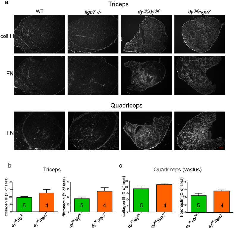
Figure 5. Fibrotic lesions are equally abundant in muscles from dy3K/dy3K and dy3K/itga7 mice.
(a) Immunofluorescence with antibodies against collagen III (coll III) and fibronectin (FN) demonstrate extensive production of fibrotic proteins in dy3K/dy3K and dy3K/itga7 muscles. (b) Fibronectin and collagen III deposition was not statistically different between these two groups in triceps (p = 0.0635 and p = 0.0635, respectively; Mann-Whitney) and quadriceps (vastus intermedius) (p = 0.1905 and p = 0.1905, respectively; Mann-Whitney). Yet, the p values for triceps muscle could indicate a trend for slightly larger areas of collagen III and fibronectin in dy3K/itga7 triceps in comparison with dy3K/dy3K specimens. The numbers of animals used are indicated in the graph. Scale bar, 300 μm.

