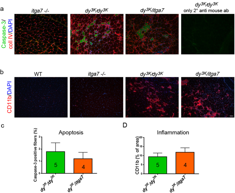
Figure 6. Muscles from dy3K/dy3K and dy3K/itga7 are equally affected with apoptosis and inflammation.
(a) Caspase-3 immunostaining (green) reveals spread apoptotic fibers or group of dying cells in both dy3K/dy3K and dy3K/itga7 muscles. Collagen IV antibody (red staining) and DAPI (blue) were used to delineate muscle fibers and show nuclei. (b) CD11b staining (red) showed infiltration of macrophages into dy3K/dy3K and dy3K/itga7 dystrophic triceps muscle. DAPI (blue) depicts cell nuclei. (c) Percentage of apoptotic fibers was not increased in double knockout triceps muscle compared to dy3K/dy3K triceps (p = 0.6828, Mann-Whitney test). (d) Amount of macrophages was not significantly different between dy3K/dy3K and dy3K/itga7 triceps (p = 0.7143, Mann-Whitney test). The numbers of animals used are indicated in the graph. Scale bar, 50 μm.

