
Figure 1.
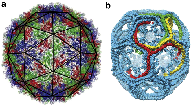
Images of (a) a viral shell (enterovirus 71) and (b) a small clathrin coat. The viral shell is a T = 3 icosahedral virus, constructed of 180 identical protein subunits. The proteins sit in three color-coded quasi-equivalent positions (grouped into triangles as seen with the dark outline in subfigure a). The clathrin coat (b) has a D6 dihedral symmetry, and is often referred to as a D6 barrel. The clathrin molecules have a triskelion (three-legged) shape that binds antiparallel leg-to-leg. The symmetries displayed by these shells allow for very detailed imaging through the technique of spatial averaging. To see this figure in color, go online.