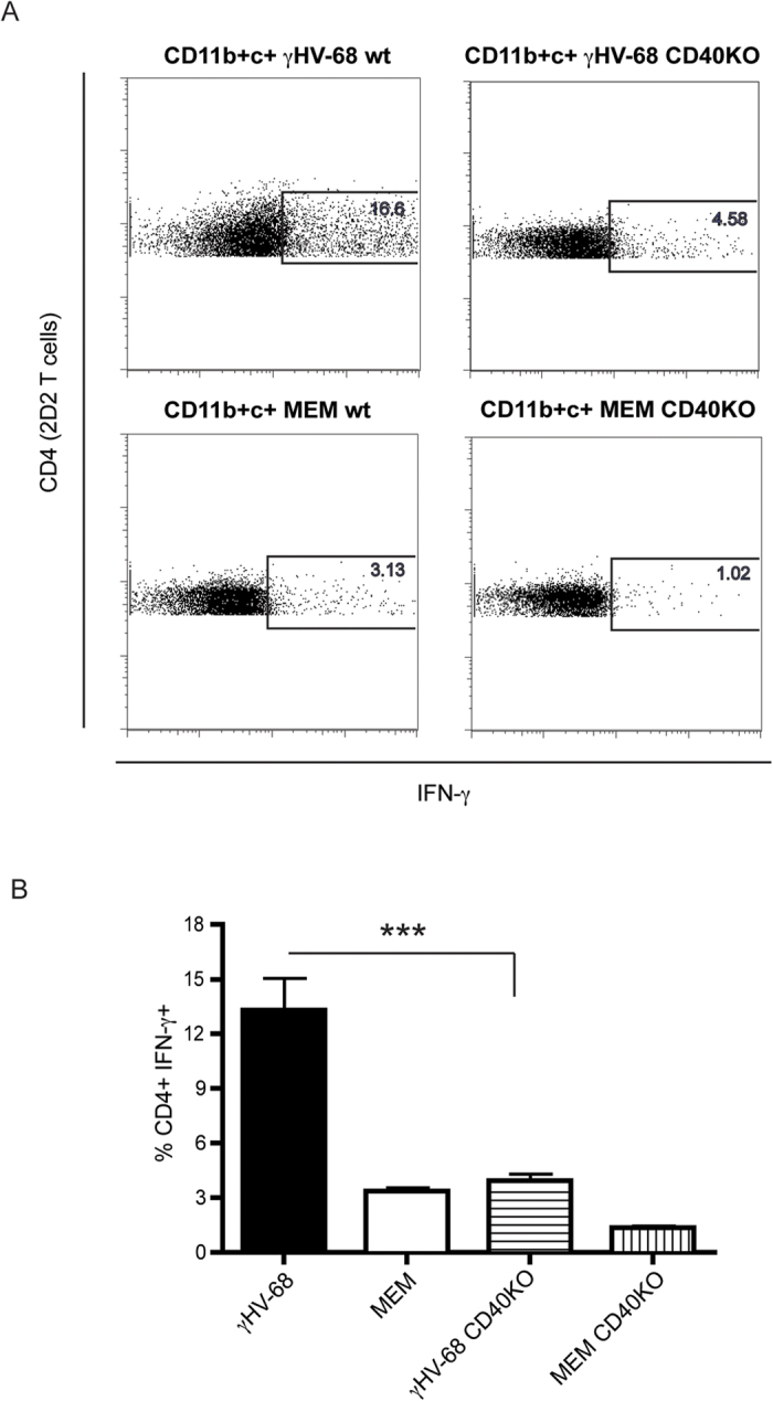
Figure 6. CD11b+ CD11c+ cells isolated from CD40KO γHV-68 EAE mice loose the ability to prime an enhanced IFN-γ response in 2D2 CD4+ T cells in vitro.

C57Bl/6 wild type and C57Bl/6 CD40KO mice were infected with γHV-68 or MEM only. Five weeks p.i., EAE was induced. At day 4 post EAE induction, spleens and lymph nodes were harvested and CD11b + CD11c+ cells were isolated. CD4 T cells from 2D2 mice were isolated at the same time. CD11b + CD11c+ were incubated with 2D2 CD4 T cells and MOG peptide for 72 hours. T cells were restimulated and stained to assess the production of IFN-γ (A) Representative FACS plot showing IFN-γ production in 2D2 CD4 T cells after incubation with CD11b + CD11c+ cells isolated from a γHV-68 wt EAE mouse (upper left panel), or a γHV-68 CD40KO EAE mouse (upper right panel), or a naïve wt EAE mouse (lower left panel) or a naïve CD40KO EAE mouse (lower right panel). (B) Histogram showing the percentages of 2D2 CD4 T cells producing IFN-γ after incubation with CD11b+CD11c+ cells from a γHV-68 wt EAE mouse (black bar), or a naïve wt EAE mouse (open bar), or a γHV-68 CD40KO EAE mouse (horizontal lines bar), or a naïve CD40KO EAE mouse (vertical lines bar). Two separate experiments with triplicate wells for each group. Data are represented as mean, error bars are SEM and were analyzed with t-test (comparing γHV-68 with γHV-68 CD40KO): ***p < 0.001.
