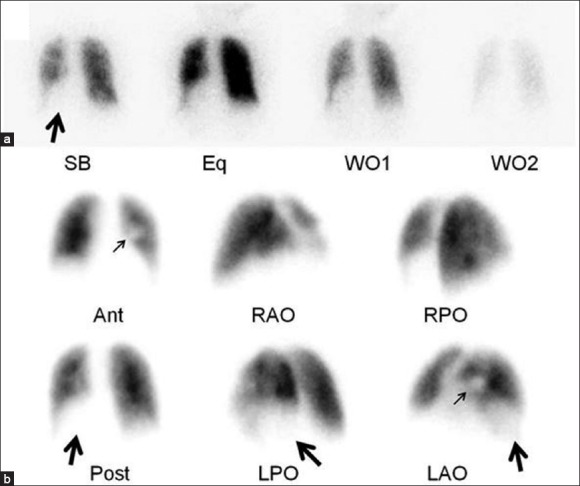
Figure 1.

Scintigraphic images from a ventilation/perfusion (V/Q) scan. (a) Dynamic ventilation portion of the exam performed with Xenon-133, which demonstrates the ventilation defect in the left lower lobe (large arrow). (b) Corresponding perfusion portion of the exam which shows the matched defects involving virtually all of the basal segments of the left lower lobe (large arrows) as well as the moderate perfusion defect in the medial portion of the left middle lung zone (small arrows) that was secondary to the known tumor
