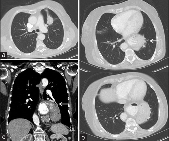
Figure 2.

Contrast-enhanced computed tomography (CT) images from the same patient. (a) Transaxial CT image in a lung window near the level of the carina shows the known left upper lobe tumor. (b) A set of transaxial CT images in a lung window at the level of the left lower lobe which demonstrates the hiatal hernia narrowing the adjacent airway and vessels (white arrow). (c) A coronal reformatted CT image in soft tissue window that also shows the compression of the left lower lobe basal segment airway and vasculature (white arrow)
