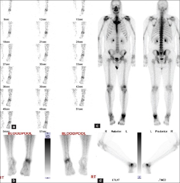
Figure 2.

Technetium-99m methyl diphosphonate bone scan images in the blood flow phase (a), blood pool phase (b), delayed whole body (c) and delayed lateral spot view of left ankle (d). Increased tracer distribution and localization are noted in blood flow and blood pool phases respectively. Delayed images demonstrated increased tracer uptake in talocalcaneal region
