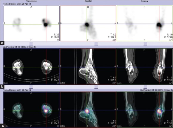
Figure 3.

Single photon emission computed tomography (a) computed tomography (b) and combined Single photon emission computed tomography/computed tomography (c) images of the left ankle in three orthogonal planes confirmed that increased tracer uptake clearly corresponds to the posterior aspect of talus, the os trigonum and their synchondrosis
