
Figure 1.
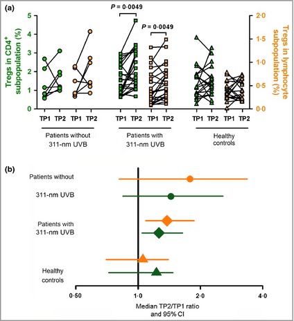
Photohardening treatment increases the number of Tregs in patients with PLE. Peripheral blood mononuclear cells of patients and healthy controls were stained with antibodies for CD4, CD127, CD25 and FoxP3. (a) Percentages of Tregs as a proportion of CD4+ cells (green symbols) or all lymphocytes (orange symbols) in PLE patients with or without 311‐nm UVB compared with healthy individuals at TP1 and TP2. (b) Median TP2 : TP1 ratio (± 95% CI) for the percentage of Tregs in CD4+ (green symbols) and lymphocyte (orange symbols) subpopulations from each of the three subject groups. Datasets were logarithmized before calculation of ratios. Patients without 311‐nm UVB, n = 7; patients with 311‐nm UVB, n = 23; healthy controls, n = 19. P‐values were determined by Wilcoxon test. CI, confidence interval; PLE, polymorphic light eruption; TP1, time point 1 (before phototherapy); TP2, time point 2 (after phototherapy); Tregs, regulatory T cells; UVB, ultraviolet B.