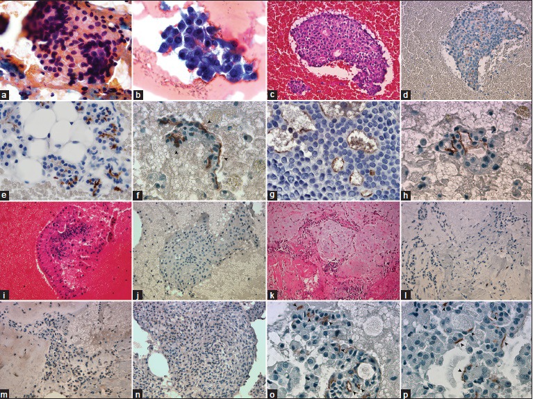
Figure 1.

(a) Large granular cytoplasm with round eccentrically located nuclei (PAP, ×1000). (b) Clusters are composed of malignant acinar cells, thin-granular large cytoplasm, and mildly atypical nuclei (Diff-Quik, ×1000). (c) Cell block of acinic cell carcinoma (H and E, ×400). (d) DOG1-K9 clone shows diffuse apical-luminal expression in acinic cell carcinoma (Immunohistochemistry, ×400). (e) Apical-luminal staining (2+) in normal serous acinar cells (Immunohistochemistry, ×1000). (f-h) Apical-luminal staining in acinic cell carcinomas, (1+, 2+, 3+), respectively, (Immunohistochemistry, ×1000). (i) Warthin Tumor is easily recognized with mature lymphocytes wrapping oncocytic cells (H and E, ×400). (k-m) Cell block of pleomorphic adenoma, areas of focal weak cytoplasmic granular positivity, and negativity in the identical case (H and E and Immunohistochemistry, ×400). (n) Focal, weak cytoplasmic granular staining in myoepithelioma case (Immunohistochemistry, ×400). (o and p) Apical-luminal staining (subcellular localization) in acinic cell carcinomas, (Immunohistochemistry, ×1000)
