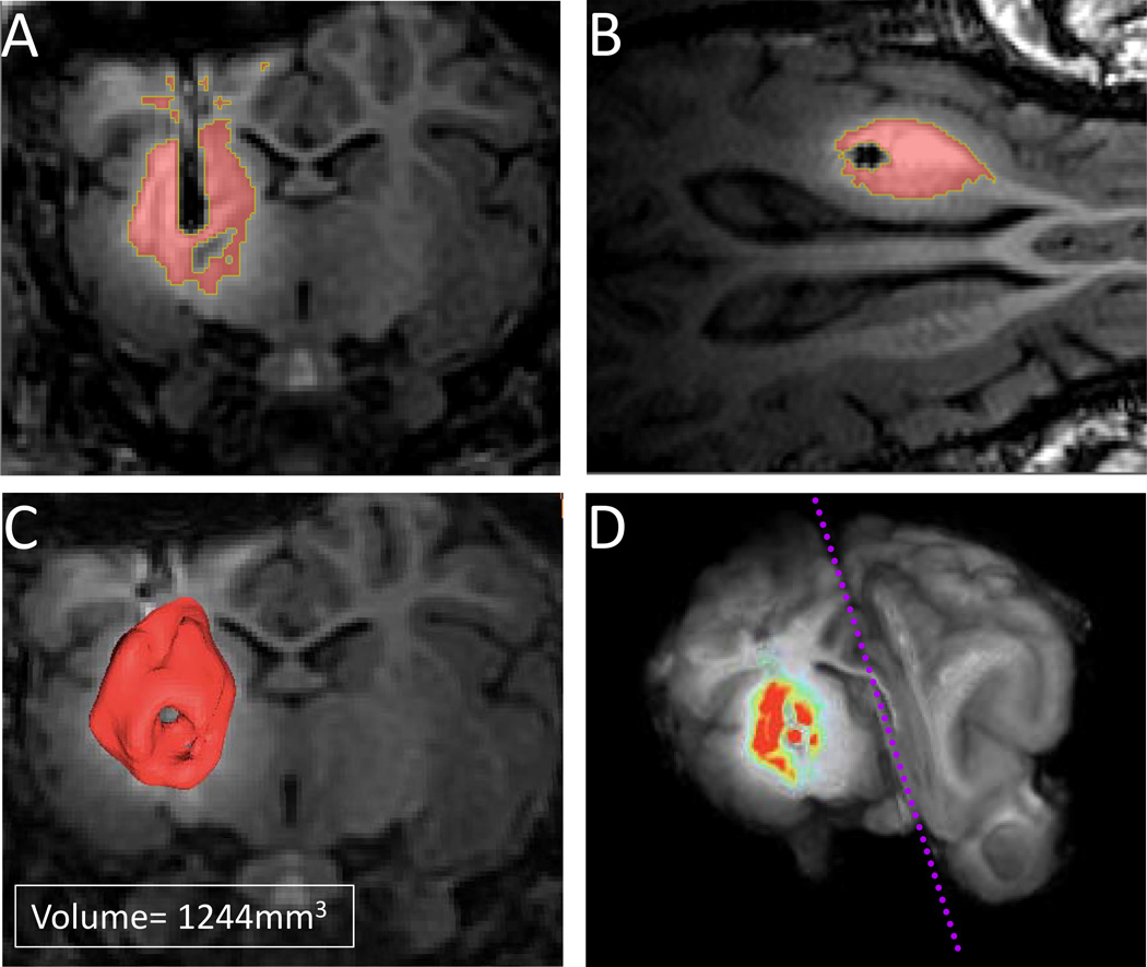
Figure 4.
A) Coronal view of the Magnevist® infusion in a subject at postoperative day 10 after 3 days of Magnevist® infusion. The volumes demonstrate the pattern of drug spread from the injection area. The entire putamen region was covered in all subjects with an average volume of average volume (1544±420mm3); B) axial view of the Magnevist® infusion at postoperative day 10 after 3 days of Magnevist® infusion; C) 3D-rendered contrast infusion in a representative subject; D) pseudocolored contrast infusion superimposed on a 3D pig brain.

