Fig. 2.

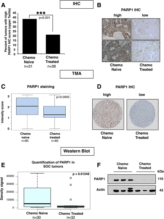
Chemo-treated tumors have lower levels of PARP1 protein than chemo-naïve tumors. a We used 69 HGSC tumors for immunohistochemistry (IHC) staining of PARP1 (training cohort). Each slide score is the product of the percentage of stained tumors cells and the intensity of the staining as described in the “Methods” section. The bar graph represents the percentage of slides with scores in the upper tertile from chemo-naïve and chemo-treated samples. Chi-square test p < 0.001. b Representative photos of PARP1 IHC from two chemo-naïve tumors and two chemo-treated tumors (20×). c Boxplot showing compilation of PARP1 intensity of staining score for the tissue microarray (TMA). The p-value tested whether there was a significant difference in PARP1 staining intensity between the chemo-naïve and chemo-treated tumors and was calculated using a two-tailed Mann–Whitney test. d Representative TMA cores stain with PARP1 antibody. e PARP1 protein levels in 62 serous ovarian cancer (SOC) tumors (30 chemo-naïve and 32 chemo-treated) were quantified with ImageJ and the density signals obtained were used to generate a boxplot. Wilcox Mann–Whitney test gave p = 0.01246. f Representative western blots of PARP1 in three chemo-naïve tumors and three chemo-treated tumors from cohort shown in e
