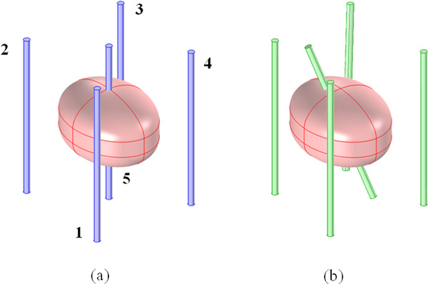
Figure 2.

3D model of the tumor in breast tissue treated with a 5 electrodes configuration. The two different configurations are reported: (a) "Symmetrical" model (blue straight standing electrodes equally spaces) and (b) "Asymmetrical" model (green electrodes) the central electrode is tilted of α angle (25°), the external electrodes are moved with displacements along the line that connect the external with the central one of about 1.8 mm, 2.8 mm and 2.1 mm for the pairs 1-5, 2-5 and 3-5 respectively. The position for the electrode number 4 remains unchanged in the two electrode configurations.
