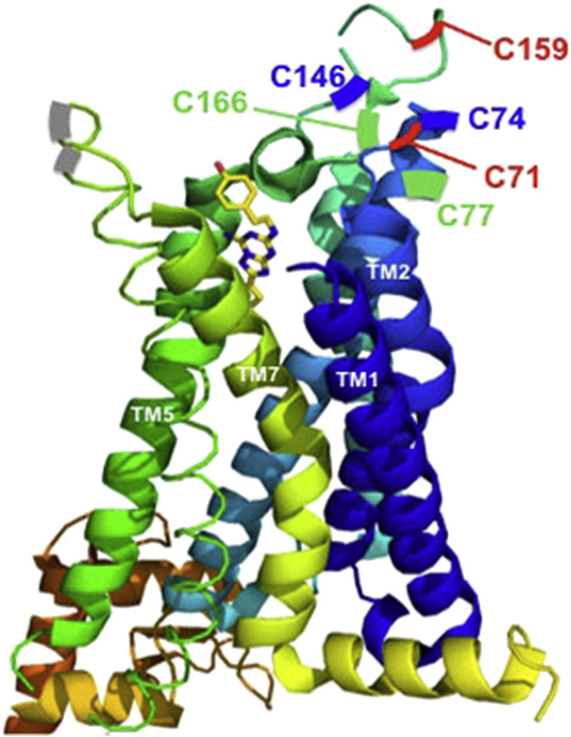
Fig. 1.
Crystal structure of A2AR bound to an antagonist, ZM 241385 [10]. The cysteines that form the disulfide bonds are color coded in green, red and blue. Adapted using PyMOL (The PyMOL Molecular Graphics System, Version 1.3 Schrödinger, LLC), Protein Data Bank identification code 3EML. (For interpretation of the references to colors in this figure legend, the reader is referred to the web version of this article.)

