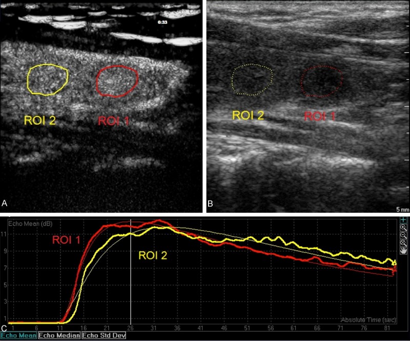
Figure 1.

Sagittal view of a solitary thyroid nodule in left lobe on double synchronous contrast-enhanced ultrasonography in a 50-year-old female patient (A and B). The nodule was solid, well margined, and wider than tall, and showed hypoechogenicity, and no calcifications (B). This nodule was proved to be benign by FNAC (Bethesda II). Region of interests (ROI) was selected in the nodule (ROI 1) and the adjacent normal thyroid tissues (ROI 2). The nodule showed rapid filling-in, rapid wash-out and hyper-enhancement as compared to the adjacent normal tissues (A and C).
