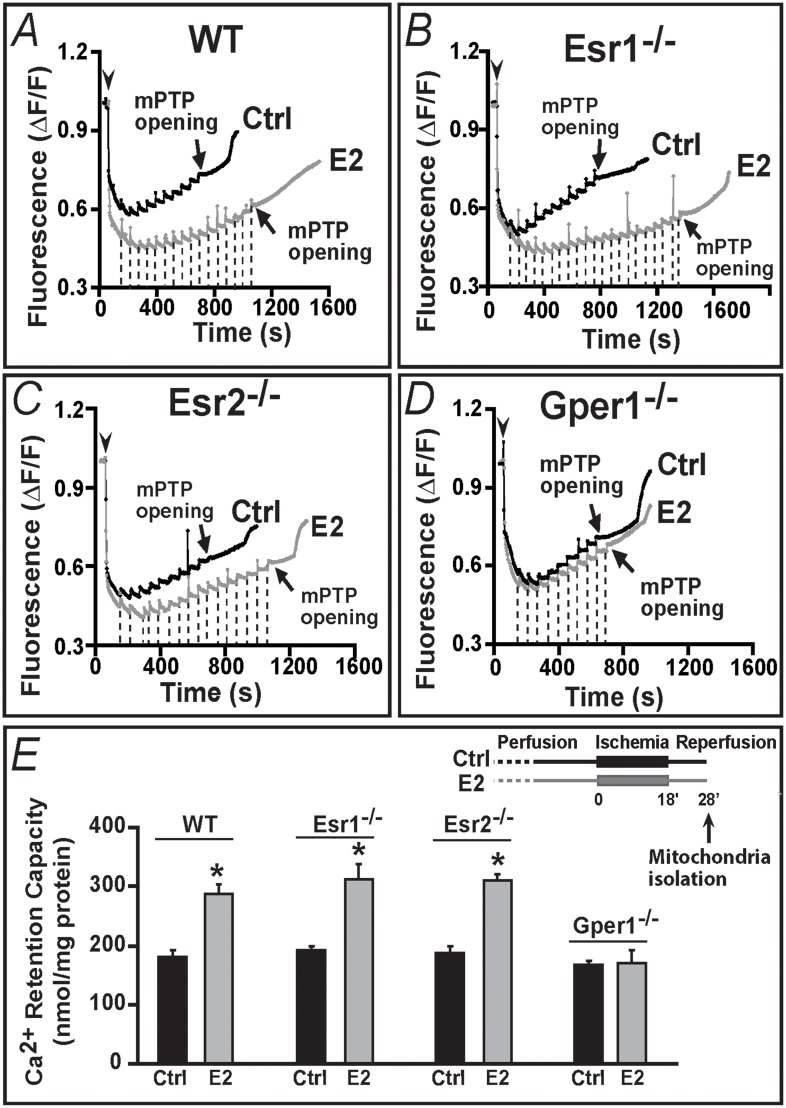
Fig 5. Gper1 activation is essential for E2 to induce increased resistance to Ca2+-overload of mitochondria from ischemic-reperfused hearts.
A-D. Spectrofluorometric recordings of Ca2+ overload in mitochondria isolated from WT (A) Esr1-/- (B), Esr2-/- (C) and Gper1-/- (D) hearts subjected to I/R (E, inset) in presence of vehicle (Ctrl, black traces) or E2 (gray traces). Arrowheads mark the time of mitochondria addition and the initial mitochondrial Ca2+ uptake. Subsequent 10 nmol Ca2+ pulses (dashed lines; only shown for E2 treated) were delivered until a spontaneous massive release was observed presumably to the opening of mPTP (arrows). Only mitochondria from Gper1-/- lost their ability to endure higher Ca2+ overload by E2 treatment (D). E. Mean Ca2+ retention capacity values (amount of Ca2+ load needed to induce mPTP opening). Inset. I/R protocol marking the time of mitochondria isolation (10 min after reperfusion begun). Values are expressed as mean±SEM; * P<0.05 control versus E2-treated groups (n = 5 hearts/group).

