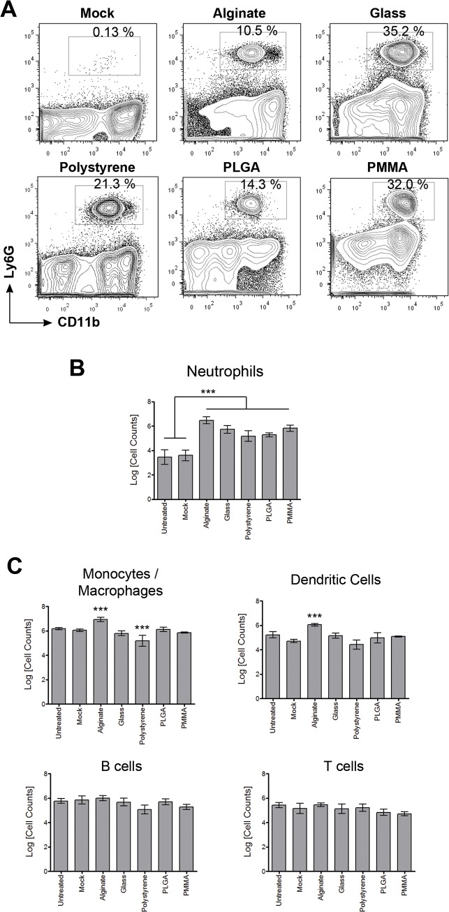
Fig 2. Increased neutrophil presence in peritoneal exudate following microcapsule implantation.
(A)–Representative flow cytometry contour plots showing percentages of neutrophils (CD11b+ Ly6G+) in the peritoneal exudate of mice implanted with microcapsules made of different materials. (B)–Counts of neutrophils in the peritoneal exudate 2 weeks following implantation of microcapsules made of different materials compared to control untreated and mock treated mice. C–Counts of monocyte/macrophage (CD11b+ Ly6G- CD11c-), dendritic cells (CD11b+ CD11c+), B cells (CD19+), and T cells (TCRβ+) in the peritoneal exudate 2 weeks following implantation of microcapsules made of different materials compared to control untreated and mock treated mice. Mock treatment entailed performing a laparotomy and injecting sterile saline (sham surgery). *** indicates p<0.001, using one-way ANOVA followed by Bonferroni post-test comparing specific sample to mock or untreated. Data are representative of at least 2 independent experiments with total n ≥ 5.

