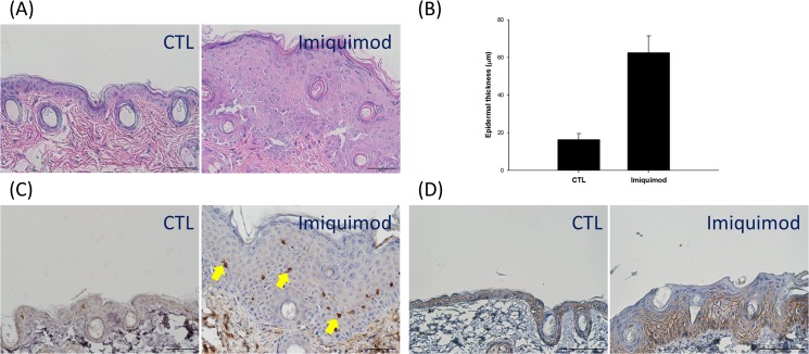
Fig 2. Histological and immunohistochemical examination of mouse skin treated with and without imiquimod cream: (A) hemoxylin and eosin (H&E) staining; (B) the epidermal thickness measured by H&E staining; (C) CD3+ T cell staining; and (D) β-catenin.
The control group corresponds to the mice treated with the vehicle.

