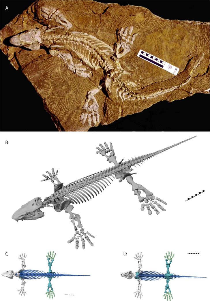
Fig 6. Digital reconstruction of Orobates pabsti (MNG 10181).
A: Holotype specimen of Orobates pabsti (MNG 10181) housed at the Museum der Natur Gotha, Stiftung Schloß Friedenstein, Germany. B: Complete digital reconstruction in a non-physiological pose. C: Dorsal aspect, D: Ventral aspect. Grey bone models were derived from highest resolution scans (≤ 75μm voxel size); green and turquoise bone models derived from lower resolution scan (≤ 150μm voxel size). Turquoise bone models were slightly re-modelled to account for partially poor visibility of bone within the matrix. Blue bone models were modelled based on superficial visibility from photos and CT-scans and according to detailed description provided by Berman et al. [17]. Shape of thoracic vertebrae was modelled after highly detailed scan of an isolated vertebra (MNG 8966). Further explanation see text.

