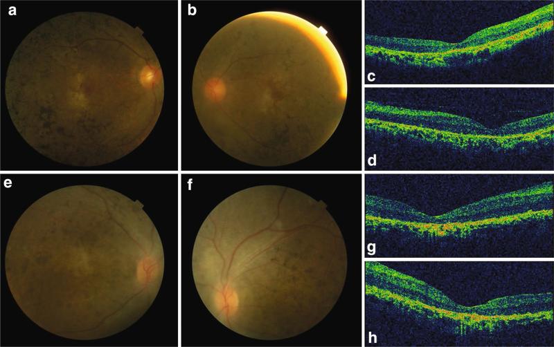
Fig. 1.
Fundus photograph and OCT images of RP patient SRF71 and LCA patient SRF92. a, b Right and left fundus pictures of SRF71. Widespread retinal bone spicule pigmentation, gold foil macular reflex and attenuation of retinal vessels were observed in both eyes. c, d Right and left eye OCT images of SRF71. The retina was thin with loss of signal of IS/OS of photoreceptor cells. e, f Right and left fundus pictures of SRF92. Widespread pigment crumbs at posterior pole and mid-peripheral area. g, h Right and left eye OCT images of SRF92. IS/OS signal was lost and the retina was atrophic

